Drug development is plagued by ineffectiveness or toxicity in significant proportions of the population. This is because blockbuster drug dose is based on the results from cumulative response curves. So the dose is decided by (for example) above 80% of the population respond. However, at this point there will be people that are under-responsive and over-responsive at this dose (figure 1). So they will get no effect or get toxicity at this dose. With certain drugs this is fatal, with 100,000 deaths a year from drug toxicity and 4 million serious adverse reaction events. Variations exist through polymorphisms and drug interactions mostly, the following will discuss some contributions to variability in drug response with some experimental examples.
Absorption, distribution and excretion
There is less variation seen in these processes than metabolism but there are some important variations that can play a big role in drug effectiveness. Absorption is generally similar in most people but disease state can cause variability, for example, 32% of patients requiring irinotecan have diarrhoea, therefore oral administration would create considerable variation with those with diarrhoea absorbing a lower dose than those without. This has to be a consideration in drug development because it suggests the drug should not be developed to be delivered via this route.
Drug distribution and excretion also become variable with disease state, distribution rate is increased if as the disease progresses hypertension and tachycardia increase. In contrast, kidney damage and failure in late stages of disease reduce the body’s ability to excrete the drug, allowing it to be active longer thus increase response.
Metabolism
Metabolism activates and inactivates drugs, so changes to metabolism will cause variation in response because reducing either of these will either lose the effectiveness of the drug or increase the action well over the usual response and be toxic. Many drugs are heavily metabolised by CYP450 enzymes and when an enzyme is metabolising one it cannot metabolise the other. This can cause variability in drug response because one person may be on the drug alone whereas anoth may be taking other drugs. This is exemplified by mibefradil which is safe alone but when administered with propranolol, inhibited CYP450 enzymes, preventing the inactivation of propranolol. A study conducted looked at patients taking both and found systolic blood pressure decreasing from over 140mmHg systolic to 60-70mmHg a fatal level. This was found to be because propranolol was not being inactivated by CYP450s and thus acting excessively on the heart. This drug was soon discontinued.
Other than drug interactions inhibiting or increasing cYP450 metabolism of another drug, CYP enzymes also get polymorphisms, relatively commonly. CYP2C9*2 is the mutant allele in 20% of Caucasians, this is a significant number of people with a loss of function of an important enzyme in drug metabolism. Testing of how much variation the presence of this allele causes showed as much as 30% differences in dose of warfarin required for efficacy. This is a large variation because if a full dose is 30% more effective it is also likely to be toxic at this level. This becomes a problem in drug development because it makes it impossible to give an accurate recommended dose and makes it likely the drug will fail in clinical trials due to toxicity. Evidence that these polymorphisms contribute heavily to variation in drug response between humans, comes from studies adding CYP2C9*2 allele to transgenic mice but using a pharmacogenetics approach of genotyping and phenotyping activity and adjusting the warfarin dose given based on the result. They found that doing this required fewer adjustments to the dose and fewer incidences of toxicity due to too high doses. This suggests that CYP polymorphisms cause lots of variations to metabolism, but pharmacogenetics can be used in drug development to get the correct dose much faster and safer and almost eliminate the variation that is seen.
The future for genetic CYP testing could be in producing transgenic mice with CYP mutations as part of animal models to identify what changes in dose are required for each polymorphism. Evidence it is accurate from measuring CYP2D6 polymorphisms to quinidine and measuring the changes in response to doses and changing dose until variability is within acceptable levels.
CYP polymorphisms are not the only causes of variation in response, other important enzymes also cause variation in response between people including thiopurine methyltransferase (TPMT). TPMT*3A-C polymorphisms cause a total loss of function of this enzyme which allows a build-up of active thiopurines this in turn leads to an increase in efficacy and toxicity which eventually leads to myelosuppression. In this case variation has been fatal, because the toxicity is quickly fatal and a full dose to some heterozygote mutants is 50% higher than the dose should be and 15% of people are at least heterozygous for a TPMT mutation. Again the relationship with drug development is that drug cannot be developed to suit most but not all because it can be fatal. This could lead to a loss of millions at clinical trials or even more if it has to be discontinued. This is more evidence that variation in patients has to be accounted for in drug development, therefore pharmacogenetics may be the approach in rug development to create more individualised medicines from drugs that are technically block busters, it should increase success in clinical trials along with reducing variation.
Non-metabolic genetics
Aside from metabolism genetics comes into other variations between patients. The same cancer can have different responses to the same drug in different people. This is also a type of variation in response. Also taken into account is the stage of cancer because as the cancer progresses mutations are random so the same cancer can look very different by stage 4. A good example of variation to the drug is the drug Herceptin for breast cancer. It acts on the HCN2 receptor and is very effective but only in 25% of cases because men do not have the receptor and the receptor can be down regulated early in the cancer. This shows the variation in drug response because the drug is very effective at treating breast cancer, but the receptor is not present in all people or all breast cancers. This causes a problem in drug development because it is obvious the drug is very effective but getting significant results is difficult because of how variable cancers are.
In summary, variation in drug response is seen more commonly in metabolism but absorption, distribution and excretion also lead to variation between people, mainly due to what state the disease is in. metabolism variation exists mainly through one drug changing the enzymes action on another drug or mutations to important enzymes. However, it is important to note that sometimes the genetics of the disorder are variable as in cancer.
Sunday, 5 February 2017

Sunday, 29 January 2017
Interesting slightly different post
I hope you enjoy the TPMT deficiency post, it is a typical pharmacogenetic interaction and I will post a follow on writing that will take this into a wider context and into the difficulties pharmaceutical companies have when developing new drugs.
Please give me some feedback on the two posts as if the consensus is good I will do a few more similar things that are not strictly Neuroscience.
Enjoy!
Thiopurine S-methyltransferase (TPMT) Deficiency
Introduction:
Thiopurine S-methyltransferase (TPMT) is an enzyme that catalyses the methylation of aromatic or heterocyclic thiol (sulphydryl) compounds (Peng et al., 2008).This function is ubiquitous, therefore the enzyme could be active in methylating toxic compounds as part of immunity such as against bacterial toxins. Its ubiquitous function is mimicked by its tissue expression and expression levels (Figure 1A). RNA-sequencing reveals that not only is it present in all tissues but it is present in specialised areas within those tissues (Figure 1B) and there is little variability between the expression levels in all tissues (Figure 1A and B vertical black lines show range), with the average expression around 0.55 FPKM (Figure 1A and B vertical red lines). The narrow range and ubiquitous nature of this enzyme show that TPMT has been highly conserved throughout all cell types and plays an important role in cell protection. The TPMT protein is coded for by the TPMT gene and has numerous polymorphisms which cause varying levels of loss of function of the expressed protein (Otterness et al., 1997). Interestingly, even those that are homozygous seem to live normal lives regardless of complete TPMT deficiency, unless they are treated during their life with the immunosuppressive thiopurine drugs, which in the absence of TPMT lead to extensive cell apoptosis. This report will discuss the TPMT gene and its mutations and assess the pharmacogenetic interaction between thiopurine drugs and the mutated alleles of this gene.

TPMT Genotype:
TPMT is located at position 22.3 on the P-arm of chromosome-6 (figure 2a), it is therefore autosomal so a person can be homozygous for one allele or heterozygous. In heterozygotes the two alleles will be expressed equally so the alleles are co-dominant but the majority of the global population are homozygotes (Weinshilboum and Sladek, 1980). Complete TPMT deficiency is autosomal recessive as it requires two copies of the mutant to produce low to absent function, however heterozygotes with one mutant allele will have a varying response to thiopurines. In the general population 89% are homozygous, 11% heterozygous and 0.3% homozygous mutant (Weinshilboum and Sladek, 1980). In 2011, Booth included evidence of 30 alleles (Booth et al., 2011) with relatively few mutations in total suggesting the allele differences are due to the combination of mutations rather than separate mutations. This observation is supported by genetic information from the Uniprot database which also shows a limited number of mutations (Uniprot, 2015). Despite a limited sample, Stanulla provides evidence that TPMT2 and TPMT3A-D account for 95% of TPMT deficiency (Stanulla et al., 2005), which is supported by equivalent studies (Collie-Duguid et al., 1999; McLeod et al., 1999). TPMT*3A is most common in Caucasians with Tai identifying 75% of the sample with TPMT*3A allele (Tai et al., 1996).In contrast, TPMT*3C is the most common allele in Asia (Collie-Duguid et al., 1999; Hiratsuka et al., 2000; Lu et al., 2006) and Africa (McLeod et al., 1999; Ameyaw et al., 1999). TPMT*3C is characterised by a single missense point mutation of tyrosine to cysteine at position 240 due to an adenine to guanine substitution in exon 10 (Figure 2B). TPMT*3A has this same mutation, in addition to an alanine to threonine mutation at position 154 (Figure 2B).

Protein structure and function in wildtype and mutant alleles:
TPMT is located at position 22.3 on the P-arm of chromosome-6 (figure 2a), it is therefore autosomal so a person can be homozygous for one allele or heterozygous. In heterozygotes the two alleles will be expressed equally so the alleles are co-dominant but the majority of the global population are homozygotes (Weinshilboum and Sladek, 1980). Complete TPMT deficiency is autosomal recessive as it requires two copies of the mutant to produce low to absent function, however heterozygotes with one mutant allele will have a varying response to thiopurines. In the general population 89% are homozygous, 11% heterozygous and 0.3% homozygous mutant (Weinshilboum and Sladek, 1980). In 2011, Booth included evidence of 30 alleles (Booth et al., 2011) with relatively few mutations in total suggesting the allele differences are due to the combination of mutations rather than separate mutations. This observation is supported by genetic information from the Uniprot database which also shows a limited number of mutations (Uniprot, 2015). Despite a limited sample, Stanulla provides evidence that TPMT2 and TPMT3A-D account for 95% of TPMT deficiency (Stanulla et al., 2005), which is supported by equivalent studies (Collie-Duguid et al., 1999; McLeod et al., 1999). TPMT*3A is most common in Caucasians with Tai identifying 75% of the sample with TPMT*3A allele (Tai et al., 1996).In contrast, TPMT*3C is the most common allele in Asia (Collie-Duguid et al., 1999; Hiratsuka et al., 2000; Lu et al., 2006) and Africa (McLeod et al., 1999; Ameyaw et al., 1999). TPMT*3C is characterised by a single missense point mutation of tyrosine to cysteine at position 240 due to an adenine to guanine substitution in exon 10 (Figure 2B). TPMT*3A has this same mutation, in addition to an alanine to threonine mutation at position 154 (Figure 2B).

Protein structure and function in wildtype and mutant alleles:
TPMT is a 245 amino acid protein consisting of 10 exons that form 9 beta sheets and 8 alpha helices (figure 3A) that fold to form the tertiary structure in Figure 3B. This figure also shows the binding site for adenosyl methionine and an adjacent binding site for 6-mercaptopurine from Peng’s study, identifying that adenosyl methionine forms at least seven hydrogen bonds here but 6-mercaptopurine only forms one (Peng et al., 2008). This study of the structures and binding sites concurs with the predicted function that adenosyl methionine binds as a methyl donor to the thiopurine, producing methylated thiopurine and adenosyl homocysteine in the reaction (figure 3C) (Peng et al., 2008). The same mechanism is expected for endogenous substances, however, no endogenous substances have been identified. It is possible that TPMT is a cellular defence to exogenous substances that attack through DNA integration. Evidence for this is highlighted by the toxic effects of thiopurine drugs in those with TPMT deficiency. In wildtype, TPMT inactivates prodrug thiopurines before they can be converted to active compounds (figure 3D) (Mlakar et al., 2016). Active compounds such as methylthioinosinemonophosphate inhibit the purine biosynthesis pathway which prevents DNA synthesis and repair or other active metabolites that insert into DNA forming interstrand crosslinks and single strand breaks that lead to apoptosis due to extensive changes in gene expression and DNA damage (figure 3D).
The tyrosine to cysteine mutation in TPMT*3C changes the structure because tyrosine has a large side chain that forms hydrogen bonds and Van der Waals interactions with residues on beta7, beta9 and alpha8. Mutation to cysteine means alpha8 interactions are lost, causing it to pull away (Rutherford and Daggett, 2008). This makes the thiopurine binding site more flexible so the thiopurine is exposed to solution. Making the thiopurine less likely to accept methylation and instead become unbound. In the TPMT*3A allele this structural deformation is coupled with a loss of hydrogen bonding between alanine and tyrosine, the double mutation forms a flattened protein and destabilises the protein causing it to be degraded by proteasomes (Rutherford and Daggett, 2008). Therefore has a half-life of 20 minutes compared to 11 hours in TPMT*3C allele (Rutherford and Daggett, 2008). This seems to be the major contributing factor as to why TPMT activity is almost absent in TPMT*3A homozygotes rather than changes in enzyme action which is the explanation for TPMT*3C allele loss of function.

Clinical features, treatments and diagnosis of TPMT deficiency:
Clinical features of TPMT deficiency are the same as the toxicities of the drugs, only the incidence and severity is higher in those with the disorder so fatality is higher (Dewit et al., 2010). To control the toxicity in those with the deficiency the dose is reduced by 50% for heterozygous and 90% for homozygous mutants (Coenen et al., 2015).
The main adverse effect is myelosuppression, a reduction in erythrocytes (anaemia) and leukocytes (leukopenia) due to the destruction of newly differentiated stem cells in bone marrow (Colombel et al., 2000). The crossover between efficacy and toxicity is evident here because when thiopurines are used to treat lymphoblastic leukaemia (increased level leukocyte derived cells), they act to reduce the number of these cells but the white blood cell loss can be excessive, causing leukopenia. This means there is a narrow therapeutic window. In those with the deficiency, leukopenia is more severe due to the excess active metabolites.
Approaches to diagnosis include phenotype testing which surveys TPMT enzyme activity by adding thiopurines and measuring the metabolite level in a purified sample (Burnett et al., 2014). Or genotype tests which sequence the gene to identify mutations that could change TPMT activity. However, a novel mutation may not be picked up (Burnett et al., 2014).
Treatments tend to deal with the toxicities as there has been no co-agonist identified that can increase the endogenous activity of the non-mutant allele in heterozygotes. Future therapy may be to reduce the degradation rate of TPMT allowing it to have a longer action. However, the most effective way would be through inhibition of the protease pathway which is ubiquitous and heavily relied on in the body. This would likely lead to cell apoptosis due to a build-up of toxic substances, so is not a viable option until another way to reduce its degradation can be found. Myelosuppression can be managed through managing anaemia, by stimulating erythropoietin with Darbepoetin alpha and neutropenia can be rescued with granulocyte colony stimulating factors as well as prophylactic treatments of infection. There has been a suggestion that treating with thioguanine may provide a less toxic alternative to other thiopurines. This has shown to be the case for inflammatory bowel syndrome, unfortunately, the licencing has been rejected due to hepatotoxicity but a derivative with some structural changes could be a future therapy.
The tyrosine to cysteine mutation in TPMT*3C changes the structure because tyrosine has a large side chain that forms hydrogen bonds and Van der Waals interactions with residues on beta7, beta9 and alpha8. Mutation to cysteine means alpha8 interactions are lost, causing it to pull away (Rutherford and Daggett, 2008). This makes the thiopurine binding site more flexible so the thiopurine is exposed to solution. Making the thiopurine less likely to accept methylation and instead become unbound. In the TPMT*3A allele this structural deformation is coupled with a loss of hydrogen bonding between alanine and tyrosine, the double mutation forms a flattened protein and destabilises the protein causing it to be degraded by proteasomes (Rutherford and Daggett, 2008). Therefore has a half-life of 20 minutes compared to 11 hours in TPMT*3C allele (Rutherford and Daggett, 2008). This seems to be the major contributing factor as to why TPMT activity is almost absent in TPMT*3A homozygotes rather than changes in enzyme action which is the explanation for TPMT*3C allele loss of function.

Clinical features, treatments and diagnosis of TPMT deficiency:
Clinical features of TPMT deficiency are the same as the toxicities of the drugs, only the incidence and severity is higher in those with the disorder so fatality is higher (Dewit et al., 2010). To control the toxicity in those with the deficiency the dose is reduced by 50% for heterozygous and 90% for homozygous mutants (Coenen et al., 2015).
The main adverse effect is myelosuppression, a reduction in erythrocytes (anaemia) and leukocytes (leukopenia) due to the destruction of newly differentiated stem cells in bone marrow (Colombel et al., 2000). The crossover between efficacy and toxicity is evident here because when thiopurines are used to treat lymphoblastic leukaemia (increased level leukocyte derived cells), they act to reduce the number of these cells but the white blood cell loss can be excessive, causing leukopenia. This means there is a narrow therapeutic window. In those with the deficiency, leukopenia is more severe due to the excess active metabolites.
Approaches to diagnosis include phenotype testing which surveys TPMT enzyme activity by adding thiopurines and measuring the metabolite level in a purified sample (Burnett et al., 2014). Or genotype tests which sequence the gene to identify mutations that could change TPMT activity. However, a novel mutation may not be picked up (Burnett et al., 2014).
Treatments tend to deal with the toxicities as there has been no co-agonist identified that can increase the endogenous activity of the non-mutant allele in heterozygotes. Future therapy may be to reduce the degradation rate of TPMT allowing it to have a longer action. However, the most effective way would be through inhibition of the protease pathway which is ubiquitous and heavily relied on in the body. This would likely lead to cell apoptosis due to a build-up of toxic substances, so is not a viable option until another way to reduce its degradation can be found. Myelosuppression can be managed through managing anaemia, by stimulating erythropoietin with Darbepoetin alpha and neutropenia can be rescued with granulocyte colony stimulating factors as well as prophylactic treatments of infection. There has been a suggestion that treating with thioguanine may provide a less toxic alternative to other thiopurines. This has shown to be the case for inflammatory bowel syndrome, unfortunately, the licencing has been rejected due to hepatotoxicity but a derivative with some structural changes could be a future therapy.
Conclusion:
In summary, the limited treatments and genetic variation of the TPMT gene in the population highlight the need for individualised treatments especially in pharmacogenetics. The wildtype allele functions as a methylating enzyme expressed ubiquitously throughout the body and may have a role in foreign immunity as no endogenous substrates have been identified. Mutant alleles for TPMT lead to damaging effects due to an inability to reduce the cytotoxic metabolites of thiopurine prodrugs and therefore cause fatal infections through myelosuppression. TPMT*3A and C are the most common mutant alleles but TPMT*3A disrupts function primarily due to an increased degradation rate, whereas TPMT*3C has a lack of function due to a change in protein structure. The negative impacts of this drug to mutant interaction are severe but their benefit outweighs the negatives because the majority of the population do not have any mutation and they are used in the treatment of conditions that are fatal.
Wednesday, 25 January 2017
Sorry about the absence
I know it has been a few months that I have been absent, however it was due to exam revision for January exams and the actually process of taking those exams.
I have a few things lined up for the people that read this blog including an interesting and slightly different post on inheritance and how mutations in metabolic enzymes can be a big problem in the drug development industry. It will begin with a post based on inheritance and then a subsequent post will delve further into its effects in pharmacology.
I am also going to release a few things specifically on neuroscience, which I know most of you like or you wouldn't be reading this.
And for those of you who are students or aspiring students, I am going to keep a short account/diary thing of my dissertation that I will publish on here. As I am doing a laboratory project not a traditional critical review so I will try to talk about lab life and how it combines with writing a dissertation, which will hopefully help some people decide what they want to do when you get here. (it will most likely apply to other biomedical sciences too so do not think it is just neuroscience labs)
(Also any of you that can, please press the follow button on my blog, I know quite a few of you people out there read it but no-one has followed it so it looks like I am talking to myself).
Thanks
I have a few things lined up for the people that read this blog including an interesting and slightly different post on inheritance and how mutations in metabolic enzymes can be a big problem in the drug development industry. It will begin with a post based on inheritance and then a subsequent post will delve further into its effects in pharmacology.
I am also going to release a few things specifically on neuroscience, which I know most of you like or you wouldn't be reading this.
And for those of you who are students or aspiring students, I am going to keep a short account/diary thing of my dissertation that I will publish on here. As I am doing a laboratory project not a traditional critical review so I will try to talk about lab life and how it combines with writing a dissertation, which will hopefully help some people decide what they want to do when you get here. (it will most likely apply to other biomedical sciences too so do not think it is just neuroscience labs)
(Also any of you that can, please press the follow button on my blog, I know quite a few of you people out there read it but no-one has followed it so it looks like I am talking to myself).
Thanks
Sunday, 20 November 2016
Experimental design: getting from cloning a new gene to finding out its role
You have cloned a new gene X. Patch clamp recording from
HEK293 cells overexpressing X revealed that X-transfected cells express large
ionic currents when these cells are mechanically stimulated (i.e. by touching
the cell with glass rod); no such currents were seen in untransfected cells.
Immunostaining of rat tissues with antibodies against X revealed that X is
highly expressed in a subset of large dorsal root ganglia and trigeminal
ganglia neurons and also in the auditory hear cells. Design the experimental
strategy to test if gene X is an ion channel, what other tissues it is expressed in and its physiological role.
Cells that express ionic currents when one recombinant gene
is implanted but no current in native cells means that this gene is likely an
ion channel. The current direction gives an indication of the role of the gene
as a large downward inflection would suggest a role in depolarisation. However,
without further experimental evidence, a whole cell patch clamp recording
cannot confirm that gene X is an ion channel. The following will discuss the
techniques for identifying whether it is an ion channel, tissue expression and
the function of the X protein.
Is it an ion channel
and what does it conduct?
Before adding the recombinant channel, a green fluorescence
protein tag sequence can be added to the gene. This would allow viewing of the
plasma membrane through Total Internal Reflection Fluorescence Microscopy (TIRF)
which emits light at an angle where the wavelengths are all reflected away from
the specimen but some photons diffuse through and excite fluorophores only on
the PM. So there will only be a strong fluorescent signal from the protein if
it is present on the PM. If this was not the case it is unlikely to be an ion
channel. Using native HEK cells as a control will make for easy comparison.
Ion channels have a distinct single-channel recording
profile so a cell-attached patch clamp to isolate one channel could be done and
record from the single channel whilst mechanically stimulating the cell. An ion
channel would provide a current trace like in figure 1.

Also this technique discounts the chance that
the gene is an ion-pump because a single pump recording is too low to be
detected. siRNA should be used to reduce the expression of the mRNA of the gene
and then redo whole-cell voltage clamp recordings and compare the first
recombinant cell recordings as a control. If the current is significantly
reduced it is likely that the gene codes an ion channel. A control using
scrambled siRNA can be used which as it has no effect on X-mRNA so if the
current still decreases then it is not due to X-mRNA knockdown as this
non-specific action is a limitation of siRNA.
Identifying the ions it conducts can begin with adding NMDG,
if the current is ablated then the channel is a cation channel. If not, it is
likely a chloride channel. A cation channel could be non-selective so the
channel can then be treated with tetrodotoxin (Na+), tetraethylammonium (K+) or
ruthenium red (Ca2+) separately, if the current is reduced by any of these then
the channel conducts that ion. If all do then it is non-selective. However, not
all subtypes of each cation channel are inhibited by these blockers, such as
NaV1.7. Therefore, the results can be confirmed by removing each cation from solution
and measuring the current.
Expression in other
tissues:
Immunohistochemistry and electrophoresis can be used in
conjunction to support the results of each test. Being as this is a newly
cloned gene it is unlikely there is an antibody for it. One could be produced
in normal animal-anti-animal technique or an epitope could be genetically
introduced into the gene of transgenic species. Tissue slices are taken and
stained with the specific primary antibody to bind and then a secondary,
fluorescent antibody. The tissue will then fluoresce under microscope if the
tissue expresses the protein. Occluding primary antibody acts as a control to
show the non-specific binding of secondary antibodies.
SDS-PAGE electrophoresis denatures and adds uniform charge
to the proteins of a sample from each tissue. The proteins are separated by
size then radio or fluorescent antibodies are used on the film which will
appear as a blot under x-ray or microscope if the protein is expressed in the
tissue. The control for this will be to normalise the expression level compared
to actin as it is expressed at similar levels in all tissues.
Testing the
physiological role of gene-X:
It is wise to use siRNA knockdown first because the siRNA
can be delivered to the dorsal root ganglion via viral mediated injection and
reduce the expression levels of the gene so behavioural and physiological
changes can be tested. It is especially useful to do first because the same
individual can be used as the control and experimental group which reduces the
variability in response and reduces the number of animals being used at this
stage. If this stage produces positive results then knockout mice can be used
to compare.
Flanking the gene with LoxP and having KO mice only express
Cre under the NF200 promoter so the gene would only be lost in large diameter
DRG neurons. After doing so it is possible to view any behavioural changes and
because it is likely a mechanically activated ion channel, use tests such as
Von Frey filament to measure changes to innocuous touch. Post-testing, DRG
neurons can be dissected and stained to identify any changes to the neuron
physiology.
Evaluation of this
experimental design:
The methods being used are ones common to this type of research
and yield reliable, reproducible results that are easily interpreted without
the need for extensive normalisation calculations. In addition, the
experimental design that is laid out uses at least two methods for collecting
data so each will support the results from the other or present discrepancies
that can be dealt with rather than drawing false conclusions. Also, with fairly
few experiments, the tissue expression, which ion is conducted, the mechanism
by which the channel is activated (mechanical) and the function can be
identified. However, the specific way mechanical stimuli open the channel is
not identified.
Transfection of genes is only 10% so large cultures are
required for success, also GM and transgenics are expensive and have stringent
guidelines for use. TIRF is also an expensive technique. Another issue is that
low levels of expression in tissues are not detected by these methods. If
required RNA-sequencing could be used instead to detect low levels. Cysteine
linked optogenetics would be useful for identifying the channel function but
can only be used if the channel is ligand activated, as this channel is
mechanical, it cannot be used.
In summary, gene-X can be identified as an ion channel
through distinct traces when measuring single channel currents and using siRNA
to knockdown the gene to view the change in current. The ion it conducts can be
identified by removing specific ions from the solution and measuring the change
in current. Electrophoresis will show the tissues expressing gene-X and
knocking out the gene will identify the function of the gene through the
changes that occur when it is not expressed.
Wednesday, 16 November 2016
Weekend Post!!
I will be releasing another post close to/over the weekend which is something a little bit different but is often forgotten about a little bit when we are reading papers and getting information and generally trying to remember all the information thrown at us.
That is how the researchers set up an experiment to eventually reach the conclusions at the end.
Therefore I am going to post a short piece that shows a simple experimental design that gets us from having cloned a novel gene to identifying what that gene does, what it is and where it is.
I will include the question so people can attempt it and think of some other methods that could be used as well or instead.
(it is not an essay about methodology which would be boring as anything for you to read, it is a short simple idea of experimental design)
Saturday, 29 October 2016
Drug Treatments to Enhance the Cognition in Patients with Attention Deficit Hyperactivity Disorder
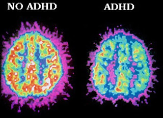
Attention deficit hyperactivity disorder (ADHD) is a cognitive deficiency characterised by problems with executive functions, specifically attentional control and inhibition. This lack of executive control leads to wide ranging effects such as a short attention span, particularly when learning and listening (Barkley, 1997). This leads to forgetfulness and a high incidence of careless mistakes. The loss of inhibition leads to hyperactivity and impulsiveness, this manifests itself as fidgeting, constant interrupting and excessive talking and movement. There are significant pathological features that are associated with ADHD, identified through functional magnetic resonance imaging and positron emission tomography. These imaging techniques (shown in figure 1) show a smaller brain overall and less brain tissue, Castellanos showed the brain to be up to 5% smaller (Castellanos et al., 1994). There are also fewer connecting fibres in the corpus callosum (Catherine, 1994), this would contribute to the difficulty in acting appropriately as there is less information passing between contralateral sensory and association areas. In addition there is evidence that the caudate nuclei are asymmetrical in a way not in concordance with those not suffering from ADHD because the right caudate has a lower volume than normal (Castellanos et al., 1994). Genetics have been implicated in ADHD and the treatments discussed later provide significant evidence that mutations in these genes play a role in ADHD development. Dopamine receptor gene for DRD4 and dopamine reuptake transporter gene for DAT1 have been shown to have polymorphisms in ADHD, treating with methylphenidate relieves symptoms by increasing dopamine present (Thapar et al., 2013). The same applies for the serotonin reuptake
transporter 5HTT and receptor HTR1B (Thapar et al., 2013) suggesting gene mutations are the main
contributing factors in ADHD. Premature birth and brain damage, both pre and
postnatally also lead to ADHD.


Dopamine is a modulatory neurotransmitter involved primarily in inhibition so methylphenidate restoring adequate dopamine to the mesocortex returns the ability to attend to stimuli. It also causes increased inhibition in the prefrontal cortex, this improves the persons ability to filter out any distractions and inhibit inappropriate behaviours and therefore enhances cognition to within normal range (HuntMD, 2006). Motor problems such as fidgeting are also inhibited through increased dopamine in the nigrostriatal pathway.
In a healthy person, methylphenidate will overstimulate dopamine receptors and cause schizophrenia-like symptoms, such as catatonia from too much motor inhibition and delusions and hallucinations (Chaudhury, 2010) therefore diagnosing ADHD must be accurate. In ADHD patients, these schizophrenia-like symptoms occur mainly in overdose but can appear at therapeutic doses (Mosholder et al., 2009), but more common adverse effects are vomiting, headaches and tachycardia (Wishart et al., 2006).
Other stimulants include Dexamphetamine and Lisdexamfetamine, these are amphetamine stimulants that are used in ADHD when methylphenidate has been contraindicated. Lisdexamfetamine is the inactive prodrug administration of dexamphetamine. They act by reversing all monoamine reuptake transporters compared to methylphenidate which is selective for noradrenalin and dopamine (Wallace, 2012). These drugs also increase the release of monoamines by increasing the activity of vesicular monoamine transporter 2 (VMAT2) which is an antiporter, moving monoamines into vesicles in exchange for protons, as well as inhibiting monoamine oxidase to prolong the action of monoamines (Wallace, 2012). These amphetamines improve function in the right caudate nucleus and prefrontal cortex, benefiting both motor and cognitive deficits (Spencer et al., 2013). They show clinical efficacy in a similar number of cases to methylphenidate (Parker et al., 2013), However, are used less frequently due to worse adverse effects, including mood swings, aggression and hyper-excitability (Punja et al., 2012).


Mirtazapine is not the only atypical antidepressant to be used to treat ADHD. Atomoxetine is a noradrenalin reuptake inhibitor used in the treatment of depression. In addition, it is also licenced as a second line treatment for refractory ADHD, when stimulants have been contraindicated or ineffective (Ghuman and Hutchison, 2014). For example, it is used in those at risk of amphetamine addiction because it has a low potential for abuse (McDonagh et al., 2011). Atomoxetine has similar effectiveness as mirtazapine at 40% (Ghuman and Hutchison, 2014) however, there are less sedative effects and no abrupt withdrawal effects (McDonagh et al., 2011) so is a safer drug for children. Additionally, it has fewer interactions with alcohol and other drugs than mirtazapine. The precise mechanism of action of atomoxetine is unclear but it elicits most of its effects in the frontal cortex and nucleus accumbens on presynaptic noradrenalin transporters.

Stimulants
as an ADHD treatment
Methylphenidate is a potent stimulant, known by the brand name Ritalin©, it is effective in 80% of ADHD patients and shown to noticeably improve symptoms. It is a noradrenalin-dopamine reuptake inhibitor that acts most potently on dopamine reuptake membrane transporters to block them. Methylphenidate has a pharmacophore (figure 2A) that binds to DATs at one site and binds in three ways (Volz, 2008). The nitrogen from the amine forms a hydrogen bond, the ester bond forms one or two hydrogen bonds with arginine on DAT and the phenyl ring forms a hydrophobic interaction by slotting into a hydrophobic binding pocket on DAT (Volz, 2008). Figure 2B shows dopamine entering between transmembrane domains 4 and 5 of the DAT1 transporter, this causes a conformational change which releases dopamine into the cell, binding of methylphenidate changes the confirmation of the transporter such that dopamine can no longer pass through the transporter. This prevents the reuptake of dopamine back into the presynaptic neuron, this means the concentration stays high enough to act on post synaptic receptors for a prolonged period of time.

Dopamine is a modulatory neurotransmitter involved primarily in inhibition so methylphenidate restoring adequate dopamine to the mesocortex returns the ability to attend to stimuli. It also causes increased inhibition in the prefrontal cortex, this improves the persons ability to filter out any distractions and inhibit inappropriate behaviours and therefore enhances cognition to within normal range (HuntMD, 2006). Motor problems such as fidgeting are also inhibited through increased dopamine in the nigrostriatal pathway.
Other stimulants include Dexamphetamine and Lisdexamfetamine, these are amphetamine stimulants that are used in ADHD when methylphenidate has been contraindicated. Lisdexamfetamine is the inactive prodrug administration of dexamphetamine. They act by reversing all monoamine reuptake transporters compared to methylphenidate which is selective for noradrenalin and dopamine (Wallace, 2012). These drugs also increase the release of monoamines by increasing the activity of vesicular monoamine transporter 2 (VMAT2) which is an antiporter, moving monoamines into vesicles in exchange for protons, as well as inhibiting monoamine oxidase to prolong the action of monoamines (Wallace, 2012). These amphetamines improve function in the right caudate nucleus and prefrontal cortex, benefiting both motor and cognitive deficits (Spencer et al., 2013). They show clinical efficacy in a similar number of cases to methylphenidate (Parker et al., 2013), However, are used less frequently due to worse adverse effects, including mood swings, aggression and hyper-excitability (Punja et al., 2012).
Atypical
antidepressants as an ADHD treatment
Mirtazapine is an atypical antidepressant with a tetracyclic structure
and the overall function of increasing the action of
serotonin/5-hydroxytyptamine (5-HT) and noradrenalin. It affects ADHD by acting
against the mutations in 5-HT transporter and the HTR1B receptor mentioned
above. Mirtazapine works by blocking 5-HT2 and 3 receptor subtypes, these are
post synaptic G protein coupled receptors (GPCRs) and ion channels
respectively. Whilst simultaneously increasing 5-HT1 receptor activity. These are
pre and post synaptic GPCRs that decrease cellular responses through inhibiting the cyclic adenosine
monophosphate second messenger pathways via phosphodiesterase (Anttila and Leinonen, 2001). It has been suggested that increasing the
serotonin to bind to 5HT1 receptors decreases cellular response further, thus
reversing the emotional dysregulation in ADHD patients, such as aggression and
being antisocial (Davis and Wilde, 1996). Mirtazapine has been shown to be <40%
effective in the most effected age groups and 50% effective in adults (figure
3A). 5-HT1B and 5-HT2B receptor types have very similar structures, so
mirtazapine blocking one and activating the other is intricate. Figure 3B shows
that the binding pocket of 5-HT2B is smaller than in 5-HT1B so mirtazapine
blocks the pocket, whereas in 5-HT1B mirtazapine binds in the pocket to
modulate the receptor, still leaving space for the binding of serotonin (Davis and Wilde, 1996).
In a healthy person there is no beneficial emotional regulation and it
does not increase mood, so it only works in someone who has depression or ADHD (Schüle et al., 2002). However, whether administering to a healthy
person or ADHD patient, it elicits the same adverse effects. One of the most
significant adverse effects is serotonin syndrome, this is an excessive
increase in serotonin leading to tremors, severe hypotension and hyperthermia
which can be fatal (Boyer and Shannon, 2005). Other adverse effects include tachycardia,
headaches and extreme drowsiness.


Summary
ADHD has very significant cognitive and emotional impairment, which leads to the loss of attention and excessive motor action such as being easily distracted and fidgeting respectively. These symptoms are associated with the pathological features of a smaller brain size, significantly fewer fibres in the corpus callosum and changes in the right caudate nucleus as shown by Castellanos’s team and the Catherine studies. The treatments for ADHD focus on reversing the symptoms and not on the pathological features because little can be done about these. Methylphenidate is a dopamine, noradrenaline reuptake inhibitor that acts primarily on the DAT1 dopamine transporter to prevent dopamine re-entering the presynaptic membrane to prolong the activity of dopamine. This leads to cognitive enhancements to normal functioning. Mirtazapine increases the action of serotonin on 5-HT1, blocking 5-HT2A and 5-HT2B at the same time. The adverse effects are significant for a child, however, the benefits the drugs have on the cognitive and emotional symptoms exceed the adverse effects.
Subscribe to:
Posts (Atom)